New paper identifies a novel small molecule targeting the p75NTR transmembrane domain that blocks melanoma metastasis

Receptor transmembrane domains (TMDs) are crucially involved in relaying ligand information from extracellular to intracellular spaces and represent attractive targets for small molecule manipulation of receptor function.

Screening a library of over 8,000 drug-like compounds with an assay based on the TMD of death receptor p75NTR, we identified a novel small molecule capable of inhibiting p75NTR-mediated migration of human melanoma cells. Employing medicinal chemistry, a more potent derivative termed Np75-4A22 was identified that blocked nerve growth factor (NGF)-mediated melanoma invasion at sub-micromolar concentrations. The specific interaction of Np75-4A22 with the p75NTR TMD was confirmed by 2D NMR. Mechanistically, Np75-4A22 was found to antagonize NGF-mediated recruitment of the actin-bundling protein fascin to p75NTR, fascin association with the actin cytoskeleton and filopodia formation. Importantly, preclinical assessment of Np75-4A22 showed high oral bioavailability, low toxicity, and significant inhibition of melanoma lung invasion in mice.

These results support further development of this approach as an alternative or complementary strategy for melanoma cancer patients that do not respond to conventional chemotherapy or immune checkpoint inhibitors. 

The paper has been published in EMBO Molecular Medicine

Read the full paper HERE

New paper reveals the necessity of GDNF receptor GFRα1 for the maintenance of adult dopaminergic neurons

GFRα1 and Ret are the two necessary components of the receptor for GDNF, a neurotrophic factor discovered in the early 1990’s for its ability to support the survival of midbrain dopaminergic neurons, including those in the substantia nigra (SN) that project to the dorsal striatum (dSTR) and degenerate in Parkinson’s Disease. Several GDNF clinical trials have been conducted to date with mixed results. Despite the physiological and clinical importance of this signaling system, whether any of its components are required for the maintenance of adult SN neurons has not yet been elucidated.

In this study, we first analyzed postnatal expression patterns of Gfrα1 and Ret in the SN and established that mRNA levels peak at mouse postnatal day 15 (P15), stabilizing after P30. Using Tamoxifen-induced deletion of Gfrα1 at 3 months of age, we found that GFRα1 is required for the maintenance of a subset of adult SN dopaminergic neurons. FluoroGold tracing of SN axons from the dSTR in mutant mice revealed that ablation of GFRα1 preferentially affects the subset of ­ GFRα1-expressing neurons that project to the STR.

In addition to the well-known neuroprotective functions of GDNF/GFRα1/RET signaling, our results establish a physiological requirement of the GFRα1 component of this neurotrophic system for the continuous maintenance of SN dopaminergic neurons in the adult brain.

The paper has been published in PLOS One

Read the full paper HERE

New paper describes a single-cell transcriptomic atlas of all cell types in the brain of 5xFAD Alzheimer mice

Alzheimer’s disease (AD) is a progressive neurodegenerative disease that is a major threat to the aging population. Due to lack of effective therapy, preventive treatments are important strategies to limit AD onset and progression, of which dietary regimes have been implicated as a key factor. Diet with high fiber content is known to have beneficial effects on cognitive decline in AD. However, a global survey on microbiome and brain cell dynamics in response to high fiber intake at single-cell resolution in AD mouse models has been unavailable.

In this study, we show that dietary inulin supplementation synergized with AD progression to specifically increase the abundance of Akkermansia muciniphila in gut microbiome of 5 × Familial AD (FAD) mice. By performing single-nucleus RNA sequencing on different regions of the whole brain with three independent biological replicates, we reveal region-specific changes in the proportion of neuron, astrocyte, and granule cell subpopulations upon inulin supplementation in 5xFAD mice. In addition, we find that astrocytes have more pronounced region-specific diversity than microglia. Intriguingly, such dietary change reduces amyloid-β plaque burden and alleviates microgliosis in the forebrain region, without affecting the spatial learning and memory.

These results provide a comprehensive overview on the transcriptomic changes in individual cells of the entire mouse brain in response to high fiber intake and a resourceful foundation for future mechanistic studies on the influence of diet and gut microbiome on the brain during neurodegeneration.

The paper has been published in BMC Biology

Read the full paper HERE

New paper characterizes subcutaneous and visceral de-differentiated fat cells

The capacity of mature adipocytes to de-differentiate into fibroblast-like cells has been demonstrated in vitro and a few, rather specific in vivo conditions. A detailed comparison between de-differentiated fat (DFAT) cells and adipose stem and progenitor cells (ASPCs) from different adipose depots is yet to be conducted. Moreover, whether de-differentiation of mature adipocytes from classical subcutaneous and visceral depots occurs under physiological conditions remains unknown.

In our latest paper, we used in vitro”ceiling culture”,single cell/nucleus RNA sequencing, epigenetic analysis and genetic lineage tracing to address these questions.

We found that in vitro-derived DFAT cells have lower adipogenic potential and distinct cellular composition compared to ASPCs. In addition, DFAT cells derived from adipocytes of inguinal origin have dramatically higher adipogenic potential than DFAT cells of the epididymal origin, due in part to enhanced NF-KB signaling in the former. We also show that high-fat diet (HFD) feeding enhances DFAT cell colony formation and re-differentiation into adipocytes, while switching from HFD to chow diet (CD) only reverses their re-differentiation. Moreover, HFD deposits epigenetic changes in DFAT cells and ASPCs that are not reversed after returning to CD. Finally, combining genetic lineage tracing and single cell/ nucleus RNA sequencing, we demonstrate the existence of DFAT cells in inguinal and epididymal adipose depots in vivo, with transcriptomes resembling late-stage ASPCs.

These data uncover the cell type- and depot-specific properties of DFAT cells, as well as their plasticity in response to dietary intervention. This knowledge may shed light on their role in life style change-induced weight loss and regain.

The paper has been published in Molecular Metabolism

Read the full paper HERE

Two new papers reveal thermogenic regulation by lipids and novel adipocyte populations in renal adipose tissue

Together these results uncover a relation between sphingomyelin abundance and fine-tuning of UCP1-mediated non-shivering thermogenesis, and pave the way for studies of Peri Renal Adipose Tissue physiology and mechanisms controlling the plasticity of brown/white adipocyte phenotypes.

The papers have been published in The Journal Of Lipid Research and eLife

Read the full papers HERE and HERE

New paper defines mouse phenotypes for human ACVR1C missense variants that correlate with altered body fat distribution

Genome-wide studies have identified three missense variants in the human gene ACVR1C, encoding the TGF-β superfamily receptor ALK7, that correlate with altered waist-to-hip ratio adjusted for body mass index (WHR/BMI), a measure of body fat distribution.

In our latest paper, to move from correlation to causation and understand the effects of these variants on fat accumulation and adipose tissue function, we introduced each of the variants in the mouse Acvr1c locus and investigated metabolic phenotypes in comparison with a null mutation.

Mice carrying the I195T variant showed resistance to high fat diet (HFD)-induced obesity, increased catecholamine-induced adipose tissue lipolysis and impaired ALK7 signaling, phenocopying the null mutants. Mice with the I482V variant displayed an intermediate phenotype, with partial resistance to HFD-induced obesity, reduction in subcutaneous, but not visceral, fat mass, decreased systemic lipolysis and reduced ALK7 signaling. Surprisingly, mice carrying the N150H variant were metabolically indistinguishable from wild type under HFD, although ALK7 signaling was reduced at low ligand concentrations.

Together, these results validate ALK7 as an attractive drug target in human obesity and suggest a lower threshold for ALK7 function in humans compared to mice.

The paper has been published in Molecular Metabolism

Read the full paper HERE

New paper reveals how p75NTR interacts with RhoGDI to regulate axon growth and neuron survival

How receptors juggle their interactions with multiple downstream effectors remains poorly understood.

In our latest paper, we report that the outcome of death receptor p75NTR signaling is determined through competition of effectors for interaction with its intracellular domain, in turn dictated by the nature of the ligand. While NGF induces release of RhoGDI through recruitment of RIP2, thus decreasing RhoA activity in favor of NFkB signaling, MAG induces PKC-mediated phosphorylation of the RhoGDI N-terminus, promoting its interaction with the juxtamembrane domain of p75NTR, disengaging RIP2, and enhancing RhoA activity in detriment of NF-kB. This results in stunted neurite outgrowth and apoptosis in cerebellar granule neurons. If presented simultaneously, MAG prevails over NGF. The NMR solution structure of the complex between the RhoGDI N-terminus and p75NTR juxtamembrane domain reveals previously unknown structures of these proteins and clarifies the mechanism of p75NTR activation.

These results show how ligand-directed competition between RIP2 and RhoGDI for p75NTR engagement determine axon growth and neuron survival. Similar principles are likely at work in other receptors engaging multiple effectors and signaling pathways.

The paper has been published in EMBO Reports

Read the full paper HERE

New paper uncovers unexpected role for activin receptor ALK4 in adipose tissue hyperplasia

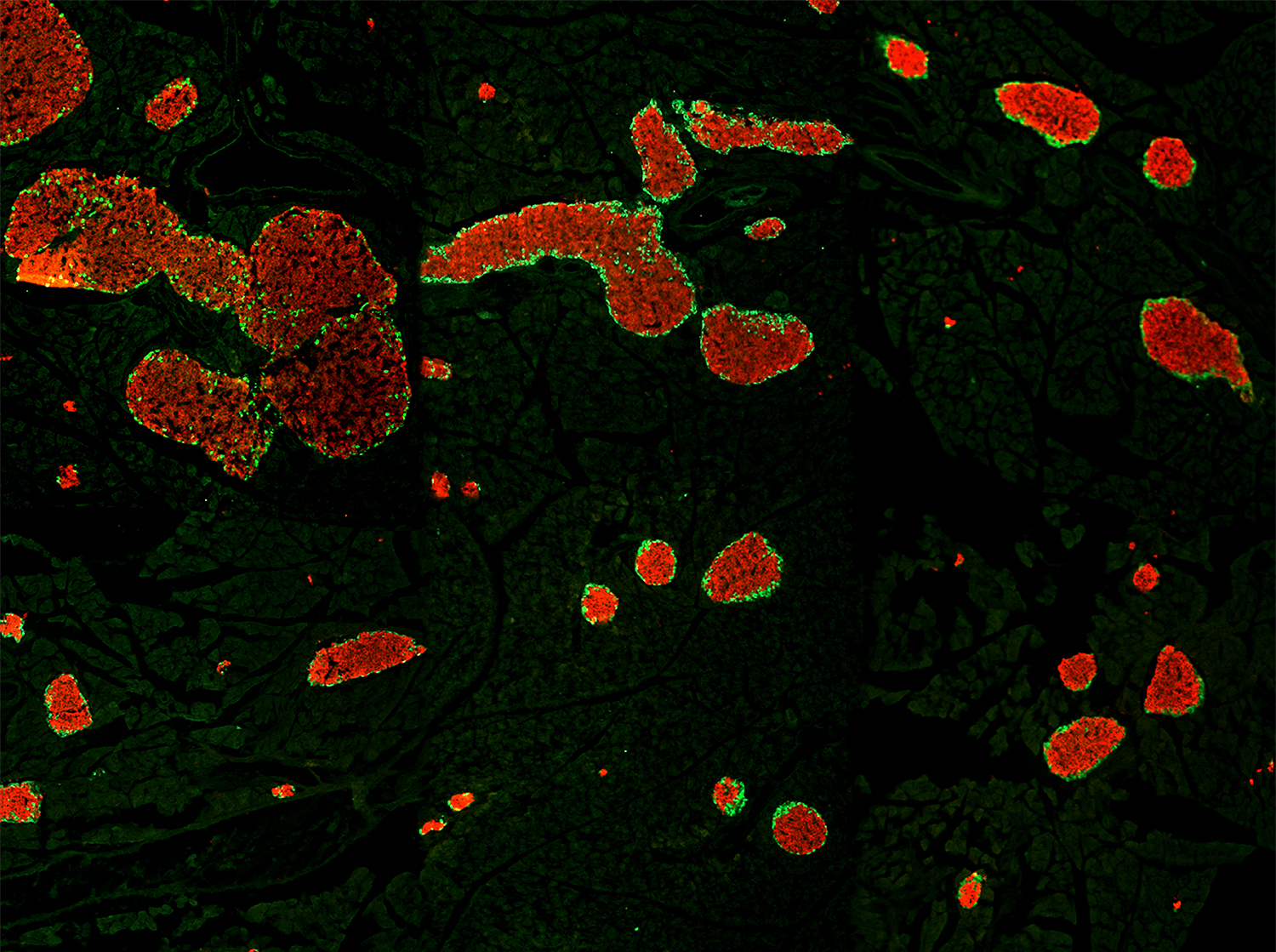
Adipocyte hyperplasia and hypertrophy are the two main processes contributing to adipose tissue expansion, yet the mechanisms that regulate and balance their involvement in obesity are incompletely understood. Activin B/GDF-3 receptor ALK7 is expressed in mature adipocytes and promotes adipocyte hypertrophy upon nutrient overload by suppressing adrenergic signaling and lipolysis. In contrast, the role of ALK4, the canonical pan-activin receptor, in adipose tissue is unknown.

In our latest paper, we report that, unlike ALK7, ALK4 is preferentially expressed in adipocyte precursors, where it suppresses differentiation, allowing proliferation and adipose tissue expansion. ALK4 expression in adipose tissue increases upon nutrient overload and positively correlates with fat depot mass and body weight, suggesting a role in adipose tissue hyperplasia during obesity. Mechanistically, ALK4 signaling suppresses expression of CEBPα and PPARγ, two master regulators of adipocyte differentiation. Conversely, ALK4 deletion enhances CEBPα/PPARγ expression and induces premature adipocyte differentiation, which can be rescued by CEBPα knockdown.

These results clarify the function of ALK4 in adipose tissue and highlight the contrasting roles of the two activin receptors in the regulation of adipocyte hyperplasia and hypertrophy during obesity.

The paper has been published in The Journal Of Biological Chemistry

Read the full paper HERE

New paper reveals how synergy between dopamine and ALK4 signaling converge to PCBP1 to control splicing of FosB and cocaine behavioral sensitization

ΔfosB is an alternatively spliced product of the FosB gene that is essential for dopamine-induced reward pathways and that acts as a master switch for addiction. However, the molecular mechanisms of its generation and regulation by dopamine signaling are unknown.

In this new paper, we report that dopamine D1 receptor signaling synergizes with the activin/ALK4/Smad3 pathway to potentiate the generation of ΔFosB mRNA in medium spiny neurons (MSNs) of the nucleus accumbens (NAc) via activation of the RNA-binding protein PCBP1, a regulator of mRNA splicing. Concurrent activation of PCBP1 and Smad3 by D1 and ALK4 signaling induced their interaction, nuclear translocation, and binding to sequences in exon-4 and intron-4 of FosB mRNA. Ablation of either ALK4 or PCBP1 in MSNs impaired ΔFosB mRNA induction and nuclear translocation of ΔFosB protein in response to repeated co-stimulation of D1 and ALK4 receptors. Finally, ALK4 is required in NAc MSNs of adult mice for behavioral sensitization to cocaine.

These findings uncover an unexpected mechanism for ΔFosB generation and drug-induced sensitization through convergent dopamine and ALK4 signaling.

The paper has been published in The EMBO Journal

Read the full paper HERE